
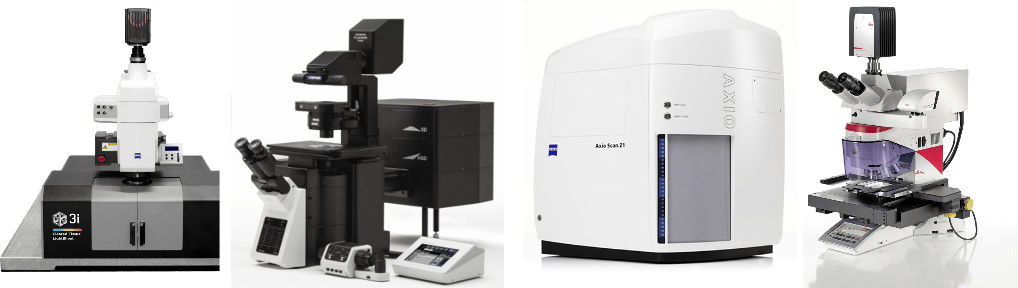
3i Cleared Tissue Light Sheet Microscope
The Cleared Tissue Light Sheet (CTLS) is a dual-side illumination lightsheet microscope for imaging whole mouse brain. It has a fully automated macro zoom microscope with high NA objectives and spatial light modulator (SLM) to optimize the light sheet for high resolution with low photobleaching.
The lightsheet microscope features:
- Lightsheet scanner with spatial light modulator, galvo scanner, beam expansion and focusing optics,
- Specimen chamber holder with motorized x,y,z translation,
- Motorized macro zoom and focus drive, filter cube turret, C-mount, touch panel controller,
- 30 fps Fusion BT sCMOS camera with 2304x2304, 6.5μm pixels,
- 3 Lasers, 488 nm, 561 nm, 637 nm,
- Apo Z 1.5/0.37 NA Objective
- SlideBook software for acquisition and Aivia Software for AI-guided image analysis.
Olympus IX-83 Inverted Confocal Laser Scanning Microscope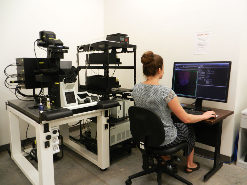
The confocal microscope allows users to acquire, process, and analyze high resolution images. Its high sensitivity and speed enables acquisition of live cell and tissue imaging. The Olympus IX-83 is a fully motorized and automated inverted imaging system. It supports fluorescence observation, high-resolution phase contrast, and long-term, time-lapse imaging.

The confocal microscope features:
- High Precision Ultrasonic Stage
- Z Drift Compensation System
- Motorized DIC Slider
- cellSens Deconvolution
- 6 Lasers, 405 nm. 488 nm, 561 nm, 640 nm, 445 nm, 514nm
- 6 Detectors
- 4x, 10x, 20x Objectives
- 40x and 100x Oil Objectives

Zeiss Axio ScanZ.1 Slide Scanner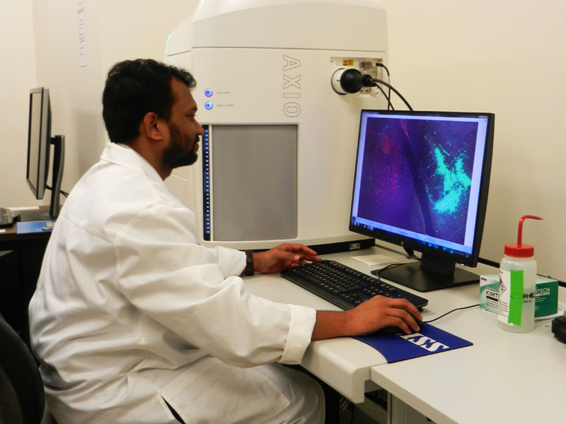
The automated slide scanner microscope allows users to digitize slides for fluorescence and brightfield. This scanner can generate a high volume of reproducible data over an extended period of time. This enclosed system scanner has 25 slide holder trays, each tray has 4 slide slots, so 100 slides can be uploaded to scan. Users can acquire up to 9 spectrally separated fluorescence channels with single band filter sets or digitize up to 5 fluorescence channels by using three filter wheels for excitation, beam splitting and emission.


Leica Microdissection System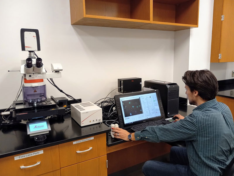
The laser microdissection microscope allows users to isolate regions of interest from subcellular structures to entire areas of tissue, both contact- and contamination-free. The user manipulates the laser location, defines the region of interest, makes the cut, and the sample drops into the collection tube. This allows for precise, analysis-ready samples.


Image Analysis Workstation
Dell workstation is a custom-built Precision 3660 tower, 13th Gen Intel® Core™ i9-13900K (36MB cache, 24 cores, 32 threads, 3.00 GHz to 5.80 GHz Turbo, 125W) processor, dual UltraSharp 27” USB-C Hub Monitor, Windows 11 Pro operating system, NVIDIA® RTX™ A4500, 20 GB GDDR6, 4 DP graphic card, and 64 GB, 2 x 32 GB, DDR5, 4400 MHz memory. Users can analyze their data using AIVIA Elevate Neuro, image visualization and analysis tools, including multiple AI-powered features, segmentation workflows, batch processing capabilities; supports a large range of applications from 2D to 5D image analysis tasks such as detection and tracking; includes Neuron Analysis recipes and the Neuron Composer to semi-automatically detect and trace neurons in 3D fluorescence data sets - as output this recipe creates soma, dendrite segments, dendrite trees, and spines.
List of the analysis software:
AIVIA Elevate Neuro, CellSens Dimension, Fiji, ImageJ, NeuronJ, NeuroGPS-Tree, Neubias-w NeuronTracing, NBLAST, CellProfiler, MATLAB, python, R studio, photoshop, Adobe Creative Cloud.
The Bio-Rad Trans-Blot Turbo Transfer System provides western blot transfers quickly and efficiently. Blot transfers occur in as little as 3 minutes and users can customize the transfer conditions.
Bio-Rad CFX96 Touch™ Real-Time PCR Detection System
This real-time PCR detection system has 6 channels, optical technology and temperature control that allows for reliable detection of singleplex or multiplex reactions.
