

Other Imaging Facilities at Tulane University
Tulane Instrumentation for Nanoscience & Innovation (TINI) : Uptown campus, Electron microscopy (TEM and SEM).
Core Facilities List at Tulane Medical school.
Biospecimen Request Form: The Biospecimen Core Laboratory (BCL) is a part of the Louisiana Cancer Research Center (LCRC) infrastructure. The BCL collects high quality samples of normal and diseased human material (e.g., whole blood, cellular blood components, bone marrow, plasma, serum, urine, benign and tumor tissue) with appropriate pathological and demographic data and to make this material available to qualified researchers.
External Funding Opportunities:
All NIH National Institute of Neurological Disorders and Stroke (NINDs) related open notices of funding opportunities (NOFOs), request for applications (RFAs), program announcements (PAs), and other NIH Guide announcements are listed.
Microscopy
- History of Microscopy: This is a link for an hour-long video lecture presented by Álvaro Tavares. An overview of the history of microscopy and simultaneously introduce some concepts in optics that are important for understanding microscopes, their components, and how it affects the resulting image.
- Microscopy Courses on iBiology: iBiology Microsocpy Series lecture videos list, including image formation, contrast, fluorescence microscopy, cameras, image analysis, and specialized microscopy.
- Lenses and Image Formation: YouTube video link for 36 min Lenses and Image formation lecture by Daniel Fletcher.
- Deconvolution: The first 20 min of the lecture about Deconvolution Microscopy by Constadina Arvanitis explains deconvolution with visual examples. Worth watching to understand deconvolution clearly.
- Olympus Confocal: This is a link to the company’s FV3000 blog page for quick access to tips and capabilities of FV3000 confocal microscope described by experts and other users.
- FV3000 Training Videos: This is a link for the list of videos provided by Olympus and includes a link to OlyVIA- software that would allow original format images to view.
- Light Sheet Microscopy: YouTube link for 30 min introduction to Light Sheet Microscopy video.
- Zeiss Axioscan Knowledge Base: Collection of short videos describing how to use Axio Scanner and Zen software, prepared by Zeiss.
- Zen Software Download: A link to download free version of the Zen software to view and export raw images.
- Leica LMD7 resources: This is a quick access link to Leica LMD7 Application website, which contains useful tips and protocols for laser capture systems.
- Super-Resolution Microscopy: What is Super-Resolution Microscopy? STED, SIM and STORM explained in an article at the Technology Networks, Neuroscience News & Research.
Image Analysis Software
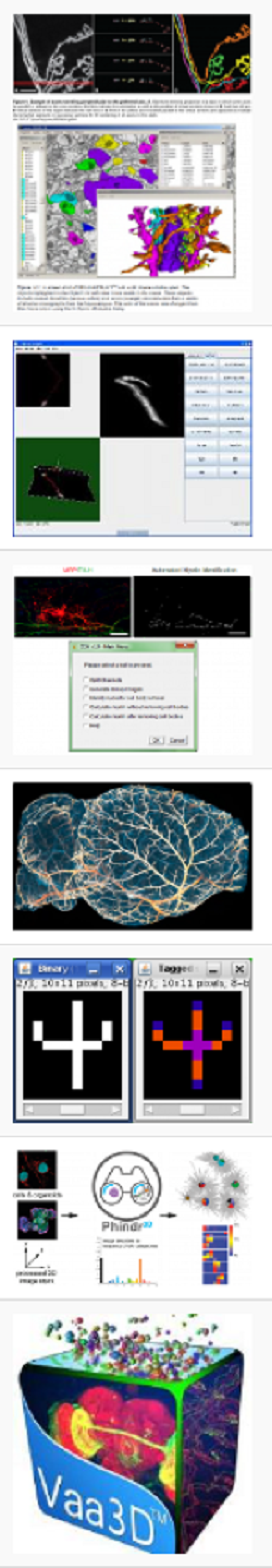
- List of software: BioImage Informatics Index website search for “neuro” related software list, where you can click on each software and find description, license status, and links to download.
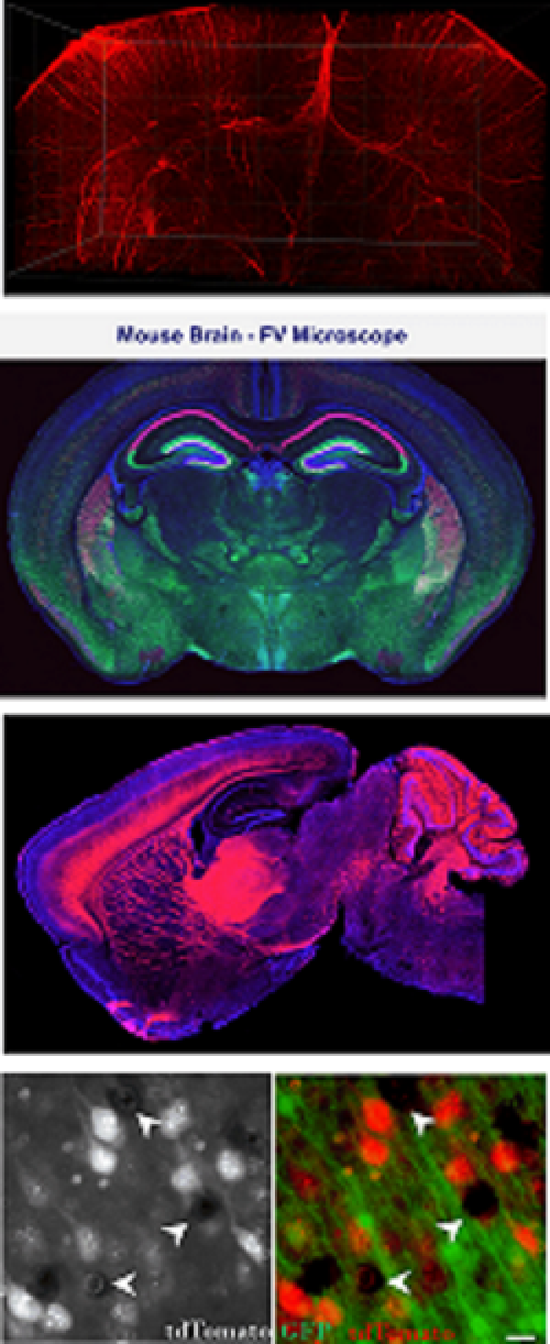
- AIVIA: is an image analysis software, which can detect, and trace fluorescence labeled dendrites, in a 3D cleared mouse brain, and classify neurons. We have a license to use in the core.
- CellSens: Imaging software is an imaging software, available to use in the confocal computer, where you can use for deconvolution and other processing, and analysis of images.
- ImageJ: links to Scientific Imaging Tutorials for the ImageJ, including download and installation.
- Cell Profilier/Cell Analyst: Free download link for the Cell Image analysis software for loading, adjusting, processing images, and exporting data.
- L-Measure: This free tool allows researchers to extract quantitative morphological measurements from neuronal reconstructions. Neuronal reconstructions are typically obtained from brightfield or fluorescence microscopy preparations using applications such as Neurolucida, Eutectic, or Neuron_Morpho, or can be synthesized via computational simulations. To get for more info click here.
- Napari is a fast, interactive, multi-dimensional image viewer, with a vibrant plugin ecosystem that expands its capability to tackle various domain-specific visualization and analysis needs. It is built on Qt (for the GUI), vispy (for performant GPU-based rendering), and the scientific Python stack (numpy, scipy, and scikit-image).Napari is an open source project on GitHub to facilitate transparency, reuse, and extensibility.It provides critical viewer features out-of-the-box, such as support for large multi-dimensional data; “layers” to simultaneously visualize images, models, and analysis results; and easy manual, interactive annotation in 3D.
- Allen Brain Map : Here on brain-map.org, you’ll find open data, analysis tools, lab resources, and information about publicly available resources.
- STAB2: (Spatio-Temporal Cell Atlas of Brain) provides a landscape of cell types as well as their regional heterogeneity and temporal dynamics across the human and mouse brain.
Basic Knowledge Resources For:
- Images: This is a link for the University of Michigan Library, describing common image information like image types, color models, image file formats, resolution, and resizing images.
- Fluorescent proteins: FPbase is a free and open-source, database for fluorescent proteins (FPs) and their properties, includes a variety of tools to: explore the lineage and directed evolution of FPs, view relationships between FP properties, compare spectra with common filters and light sources, calculate FRET relationships, create and share curated protein collections,and build a spectra viewer customized for the specifications on your microscope.
- Tissue Clearing: Collection of protocols and tissue clearing reagent information for light sheet microscopy.
- Addgene: to search for a plasmid, search addgene, a nonprofit plasmid repository.
- Protein Modifications: is a search results at the Expasy, Swiss Bioinformatics Resource Portal, which provides access to over 160 databases and software tools, from genomics, proteomics and structural biology to evolution and phylogeny, systems biology, and medical chemistry.
- Biological Information Resource: University of Pittsburg, Health Science Library System, provided an open-source online bioinformatics resource collection, links to molecular databases and software tools.
- Imaging & Microscopy Magazine: Wiley Analytical Science magazine has been providing readers with thought-provoking and relevant information on developments and trends in microscopy methods.
